


Comparison of Intraocular Cytokine Levels of Choroidal Neovascularization Secondary to Different Retinopathies
age-related macular degeneration (AMD);central serous chorioretinopathy (CSC);choroidal neovascularization;cytokine;high myopic;polypoidal choroidal vasculopathy (PCV);vascular endothelial growth factor LabEX支持文献


- Front Med (Lausanne)
- 2021
- 5.058
- 8:783178.
- Human
- luminex
- Aqueous humor
- 生物标志物
Abstract
LabEx Luminex平台助力探索脉络膜新生血管的炎症细胞因子相关机制
本周为大家带来的文献为发表于Front Med (Lausanne). (IF: 3.1)的” Comparison of Intraocular Cytokine Levels of Choroidal Neovascularization Secondary to Different Retinopathies”。本文使用了LabEx提供的Luminex检测服务。
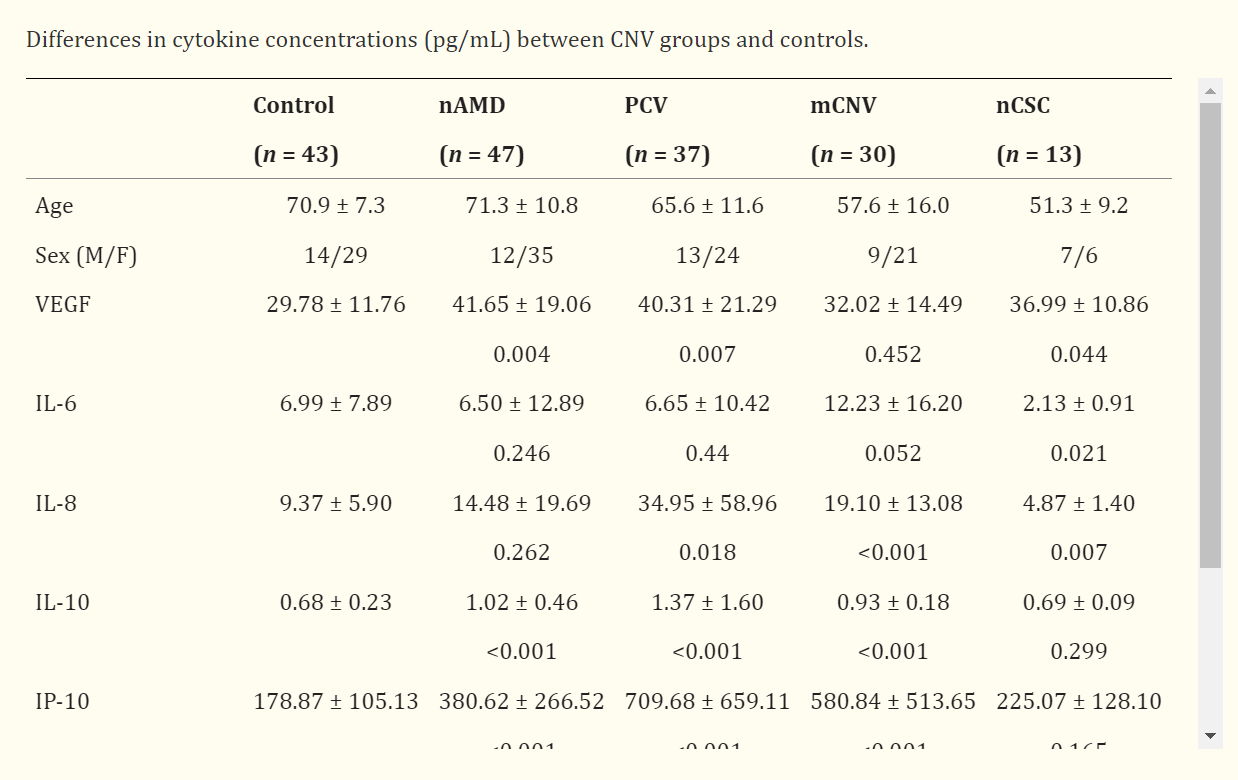
脉络膜新生血管(CNV)是脉络膜中新血管的形成。它通常发生在黄斑区,引起黄斑出血和视网膜下的浆液性渗出,从而导致失明。虽然人们对CNV的发病机理仍知之甚少,但认为它涉及多种细胞生长因子,而血管生成受促血管生成细胞因子和抗血管生成细胞因子之间动态平衡的控制。本研究调查并比较了不同 CNV 疾病和 CNV 类型中 VEGF 和其他炎症细胞因子的水浓度。此外,研究还初步探讨了血管内皮生长因子水平与光学相干断层血管成像(OCTA)图像上 CNV 大小之间的关联。
这项观察性研究包括 127 只患有 CNV 的天真眼和 43 只患有白内障的对照眼。在进行玻璃体内抗血管内皮生长因子注射或白内障手术前采集房水(AH)样本。采用多重串珠法测定多种炎症细胞因子,包括血管内皮生长因子、白细胞介素(IL)6、IL-8、IL-10、干扰素诱导蛋白 10(IP-10)和单核细胞趋化蛋白 1(MCP-1)的水平。血管生成指数定义为 IP-10 与 MCP-1 的比值。此外,还评估了AH细胞因子水平、黄斑中心厚度(CMT)和光学相干断层血管造影(OCTA)显示的CNV大小之间的关系。
LabEx提供的Luminex检测服务:
在白内障手术或玻璃体内注射抗血管内皮生长因子之前采集房水(AH)样本。使用 30 号胰岛素注射器通过角膜缘穿刺抽取约 0.05 mL 的房水样本。每份 AH 样品都被立即转移到无菌塑料管中,并在检测前保存在 -84°C 温度下。使用 Luminex 200测量未稀释的 AH 样品中血管内皮生长因子、白细胞介素(IL)6、IL-8、IL-10、干扰素诱导蛋白 10(IP-10)和单核细胞趋化蛋白 1(MCP-1)的水平。使用试剂盒提供的标准曲线计算每种细胞因子的浓度(pg/mL)。
重要发现:
除近视 CNV 组(P = 0.452)外,其他所有 CNV 组的 VEGF AH 浓度均显著高于对照组(所有比较的 P < 0.05)。除新生血管性中央浆液性脉络膜视网膜病变外,所有 CNV 病变组的 IL-8、IL-10、IP-10 和 MCP-1 水平均明显高于对照组(所有组间比较,P < 0.05)。血管生成指数在所有 CNV 疾病中均明显升高(所有比较中 P < 0.05)。血管内皮生长因子水平可能与 OCTA 上 CNV 的大小有关(P = 0.043)。


本网站销售的所有产品及服务均不得用于人类或动物之临床诊断或治疗,仅可用于工业或者科研等非医疗目的。

 沪公网安备31011502400759号
 营业执照(三证合一)

 labex@univ-bio.com
 上海市浦东新区古丹路15弄18号楼4楼
